On Call with Hilltop Bio: Virtual Rounds
On Call with Hilltop Bio: Virtual Rounds
By Dr. Chris Wickliffe & Dr. David Dutton
Edited into blog format from a live clinical discussion
From aging eventers to elite young performance horses, advances in amniotic bioscaffold technologies are reshaping how equine veterinarians manage joint disease, soft-tissue injury, foot pathology, and even difficult corneal ulcers. In a recent case discussion, Dr. Chris Wickliffe and Dr. David Dutton walked through several real-world cases highlighting how Strydaflex, Regenaflex-RT, and PureOptic are changing outcomes for horses that historically struggled with conventional therapies.
Below is a recap of some of the most compelling cases, along with practical dosing considerations and clinical timelines.
22-Year-Old Eventer With Cushing’s Disease
Cash, a 22-year-old warmblood and former eventer turned schoolmaster, had long managed age-related stifle and hock lameness with steroid therapy. After he was diagnosed with Cushing’s disease at age 18, steroids were no longer an option. Dr. Chris Wickliffe transitioned Cash to Strydaflex to maintain comfort and keep him working with his young rider.
Protocol: Strydaflex administered to both hocks and stifles every 6–9 months.
Outcome: Maintains a Grade 1 or better lameness score on flexion, continues training 4 days/week, and competes at lower levels without steroid exposure. This protocol is now used successfully in similar older, metabolically compromised horses within the practice.
Young Cutting Horse With Persistent Lower-Hock Pain
A 4-year-old cutting horse presented to Dr. David Dutton with a grade 3/5 right-hind lameness isolated to the lower hock joints. Imaging—including radiographs and ultrasound—was unremarkable. After the horse showed little improvement with the standard combination of HA, steroids, and shockwave, Dr. Dutton elected to use Strydaflex.
Protocol: Strydaflex administered to TMT/DIT.
Outcome: Returned to full soundness within days and maintained soundness long-term. A review of 18 similar cases in the practice showed 89% sound at one month and 100% sound at six months after Strydaflex treatment.
Thin-Sole, Negative-Angle Podiatry Case
A dressage gelding presented to Cascadia Equine with soreness in all four feet. Diagnostics revealed severe thin soles (0.5 cm), negative plantar angles, chronic abscessing, and wall separation. Leveraging his farrier background, Dr. Chris Wickliffe developed a multi-modal podiatry plan incorporating Regenaflex-RT and corrective shoeing.
Protocol: Regional limb perfusion using Regenaflex-RT every other month, combined with corrective farriery.
Outcome: Sole depth improved to >1.0 cm, wall thickness increased, recurrent abscessing resolved, and the horse returned to normal shoeing and regular riding after 12 months.
Deep, Melting Corneal Ulcer
A 2-year-old Quarter Horse filly was brought to Dr. Dutton’s practice with a melting corneal ulcer measuring 1 cm in diameter, with a 1 mm stromal defect reaching Descemet’s membrane. The owners reported the injury had occurred at least three days earlier.
Protocol: Atropine, triple antibiotic ointment, silver sulfadiazine, and PureOptic twice daily for 16 days. From days 16 to 45, all other medications were discontinued, while PureOptic continued to support remodeling.
Outcome: Melting halted within one week. By Day 16, neovascularization regressed and comfort returned. By Day 45, the ulcer had fully resolved with excellent remodeling.
Fetlock Wound With Fistulous Tract
Dr. David Dutton’s final case involved a 2-year-old cutting gelding with a lateral fetlock wound initially managed on the farm by capable owners. When the drainage failed to improve, the horse was brought in for evaluation. Ultrasound revealed synovial fluid leaking from the wound, confirming a fistulous tract extending to the plantar pouch of the fetlock, an issue that significantly complicated healing. Fortunately, there was no evidence of active synovitis, indicating the joint was likely contaminated but not yet infected.
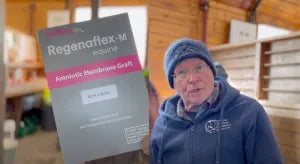
Protocol: Local perfusion of the tract with Regenaflex-RT and placement of Regenaflex-M under bandage; systemic antibiotics for 11 days.
Outcome: Drainage ceased and the tract sealed by Day 11; the wound was effectively healed by Day 18, avoiding surgical lavage and reducing overall recovery time.
Conclusion
Across joint disease, corneal ulcers, synovial wounds, and complex podiatry cases, one theme emerges: modern biologics are expanding treatment options where traditional therapies plateau or become unsafe.
For aging horses, young athletes, and fragile feet alike, products like Strydaflex, Regenaflex-RT, and PureOptic are reshaping clinical expectations and elevating what’s possible in equine performance medicine.
This content is provided for informational purposes only and represents the independent professional views of the contributing veterinarians. Hilltop Bio assumes no responsibility or liability for the accuracy or clinical application of the information presented herein.