Check Ligament Injury in Young Mare

Check Ligament Injury in Young Mare
Age: 9yo Sex: Mare Breed: KWPN Discipline: Jumper, last shown 1.15m
Treated by: Dr. Tim Ober, DVM Clinic: John R. Steele & Associates, Inc.


History
•Patient pulled left LF shoe July 10th, 2021 while in paddock, both clips left in foot
•Lame at a walk
•Client packed the foot and treated with Bute
•Patient improved slightly, but lame at trot
•Within 2 days, swelling in LF and LH, appearing like cellulitis. Swelling improving.
Exam Findings
First Exam - July 17th: patient walking sound, but 2/5 lame on LF at a trot. No elevated pulse noted. LF and LH swollen with what appeared to be cellulitis, but showed steady improvement.
Second Exam - July 19th: Mild swelling of the LF mid tendon area observed. No heat or pain on palpation noted. Patient 1/5 lame on the LF in a straight line and on a circle. Significantly more lame under tack than in hand, appearing 3+/5 lame on the circle to the right. Diagnostic block on LF was highly PDN positive with significant improvement to the right, but still showed some lameness to the left. Ultrasound showed desmitis in the LF ICL.
Treatment
July 21st: LF carpal sheath injected with Hyaluronate and Triamcinalone. Limb showed improvement for a week before starting to swell again.
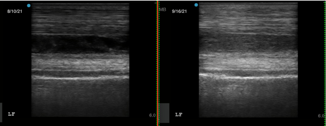
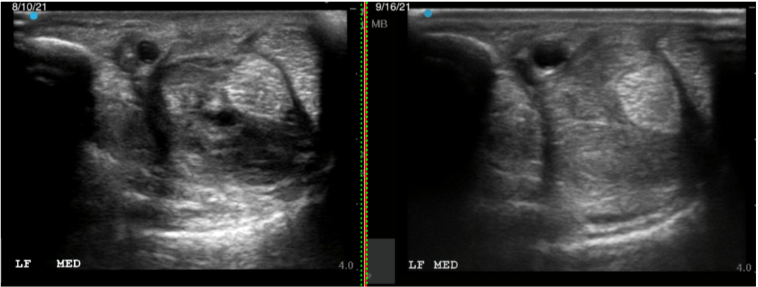
August 10th: Recheck ultrasound showed LF ICL desmitis to be more significant than on first examination. Heat and pain on palpation noted. Further treatment options were discussed.
August 27th: Regenaflex-RT administered to the LF ICL using ultrasound guided injection.
August 29th: Upon bandage removal, the LF leg appeared about 50% smaller than before the injection of the ICL. No heat or pain was observed on palpation.
September 16th: Follow-up ultrasound on 9/16/21 showed significant improvement.