Case Report: Wound Management of a Serious Canine Bite

Case Presentation
On September 3, 2025, a healthy dog presented for emergency care following a serious dog fight. The patient sustained extensive bite wounds cranial and caudal to the left ear. While the pinnae and internal ear canals remained unaffected, the surrounding tissue suffered significant trauma, including puncture wounds near the base of the ear. The primary defect measured approximately 17.5 cm x 12.5 cm (7” x 5”). Due to the location of the injuries and the amount of skin that was affected, a primary surgical closure was not possible.
 Images showing wound on case presentation. From right to left, the first two images show wound cranial to ear canal and the final image shows the wound caudal to the ear canal.
Treatment Protocol and Clinical Progress
Due to the severity of the tissue damage and the high risk of infection, a multimodal treatment plan was initiated immediately. Multiple active drains were placed both cranial and caudal to the ear to manage exudate and reduce dead space, while a course of broad-spectrum antibiotics and NSAIDs was started to manage potential infection and pain. To further support the healing process, Regenaflex-K9 was administered into the healthy tissue surrounding the wound.
Five days later during the first follow-up assessment, the attending veterinarian noted a marked decrease in inflammation. Robust granulation tissue was already evident along the wound margins, accompanied by notable wound contraction. Both the clinical team and the owner reported a visible improvement in the patient’s comfort levels, allowing for the successful removal of all surgical drains.
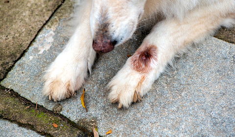
 Images showing wound 5 days after Regenaflex-K9 administrations. Images on the right and in the middle show wound cranial to the ear canal and final image shows wound caudal to ear canal.
Resolution and Long-Term Outcome
By day 24 post-treatment, the wound was nearly resolved. The initial 7” x 5” defect had contracted significantly; the region caudal to the ear was fully epithelialized, leaving only a minor residual wound cranial to the ear canal. The tissue surrounding the remaining defect displayed minimal residual inflammation and the onset of hair regrowth.
 Images taken at the final recheck, 24 days post-administration. Images on the right and in the middle show wound cranial to the ear canal and image on left shows the near total closure of wound caudal to ear.
Conclusion
This case underscores the clinical utility of regenerative devices in facilitating a healthy healing environment for severe soft-tissue injuries. The versatility of such devices provide clinicians with the necessary flexibility to adapt protocols for complex wounds that might be difficult to manage otherwise. Ultimately, this patient’s success underscores the value of a multimodal approach, demonstrating how combining standard infection control and pain management with advanced regenerative devices can enhance healing trajectories, even in cases where traditional therapies prove ineffective or impossible.